Research
In addition to scientific collaborations with other research groups, LCAM works on the development of novel microscopic techniques enable novel or enhanced contrast or resolution to boost new biological research. New microscopic techniques are being developed in close collaboration with commercial companies.
Below one can find the general fields of interest of our LCAM staff.
Functional Imaging
A main research effort is the development and application of different kinds of functional imaging microscopy. Functional imaging is a collection of microscopy techniques aimed at quantifying biomolecular interactions and cellular processes including cell signaling mechanisms. They include several modalities, e.g. Fluorescence Lifetime Imaging Microscopy (FLIM), Förster Resonance Energy Transfer techniques and Fluorescence Fluctuation techniques.
Fluorescence Fluctuation Spectroscopy (FFS)
One of the most intriguing challenges in life sciences is to understand how a complex mixture of molecular particles and structures can make up a living cell. Despite the immense number of studies still much is unknown about the molecular basis of numerous biological processes such as cell proliferation, differentiation, intra- and extra-cellular communication and apoptosis. To increase our understanding about the complexity of these processes in living cells, experimental and especially quantitative data on the spatial-temporal organization is required.  Fluorescence based techniques are ideal tools for this type of studies.
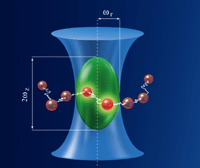
Fluorescence Fluctuation Spectroscopy (FFS) is a family of fluorescence techniques that is capable of detecting concentration, dynamics and interactions of fluorescent particles down to the single-molecule level and, if desired, in the living cell. We are applying, optimizing and expanding FFS techniques like FCCS, PCH, N&B, PIE-FLCCS, stICS and RICS to describe signal transduction pathways, like the Galpha protein and MAPK signaling pathway quantitatively. Thereto, the proteins of interest are genetically labeled with the various color- and lifetime variants of the green fluorescent protein (partly developed in our laboratory) expressed in living cells and studied by advanced fluorescence microscopes.
for more info contact Dr. Mark Hink
_
Förster/Fluorescence Energy Transfer (FRET) and Fluorescence Lifetime Imaging Microscopy (FLIM)
FRET is the phenomenon that after excitation of a donor fluorophore it transmits its excited state energy to an acceptor chromophore in its close vicinity. For common sets of donor and acceptor fluorophores (like Fluorescent Proteins, FPs) FRET only occurs within a distance of 0-10 nm. FRET is both distance and orientation-dependent and thereby it can provide direct contrast of molecular interactions and molecular conformations within living cells. FRET is manifested and can be detected with a variety of techniques. For instance, upon FRET the donor fluorescence (quantum yield/lifetime) is reduced and the acceptor fluorescence is sensitized (or enhanced). So by ratio imaging microscopy, spectral imaging microscopy, acceptor photobleaching microscopy, and by fluorescence lifetime imaging microscopy, FRET can be quantified. At LCAM-FNWI all these modalities are implemented in wide-field, confocal and TIRF setups.
FLIM is a particular quantitative FRET detection technology since it quantifies not the intensity but the duration of the excited state of the donor molecules. Hereby FLIM provides a signal that is independent of local fluorophore concentration in cells or (moderate levels of) photobleaching. FLIM is implemented in time-domain and in frequency domain, and in confocal and wide-field, TIRF and spinning disc optical setups. Detailed standardized data analysis routines are implemented aimed at quantifying lifetime reduction due to FRET, generating FRET contrast in biosensors, and at lifetime unmixing with polar plot analysis.
for more info contact Prof. Dr. Dorus Gadella, Dr. J. Goedhart, Dr. M.A. Hink or Dr. Marten Postma
_
Super-Resolution Microscopy
Recently, the Nobel Prize for Chemistry (2014) was awarded to three pioneers in the field of super-resolution microscopy. This does not only indicate that this is a fast developing and important field of science, but SR-microscopy (or “nanoscopy”) also gives spectacular sharp images revealing cells at the nano-scale. The resolution of a light microscope is limited by the wavelength of light. This resolution limit is typically around 250 nm. With new super-resolution techniques it is possible to push this limit down to 30-50 nm, however, only for cells that are fixed (and therefore dead). In our group, we focus on the development of super-resolution technology that can be used for live-cell imaging. For these live-cell super-resolution techniques we aim at resolution in the range of 100-170 nm.
It is well known that there is a spatial limit to which light can focus: approximately half of the wavelength of the light you are using. But this is not a true barrier, because this diffraction limit is only true in the far-field and localization precision can be increased with many photons and careful analysis. The image of a point source on a microscope detector is called the point-spread function (PSF), which is limited by diffraction to be approximately half the wavelength of the light. But it is possible to simply fit that PSF with a Gaussian to locate the center of the PSF, and thus the location of the fluorophore with a much higher accuracy Betzig et al. (Science) developed photo-activated localization microscopy (PALM) while Zhuang and co-workers used a similar technique called stochastic optical reconstruction microscopy (STORM).
In both techniques samples filled with many dark fluorophores are imaged. The dyes can be photoactivated into a fluorescing state by a flash of light. Because photoactivation is stochastic, only a few, well separated molecules “turn on”. Then Gaussians are fit to their PSFs in order to localize the centre of the particle. After the few bright molecules photobleach (sometimes actively by using another differently colored excitation source), the next flash of the photoactivating light activates random fluorophores again and the PSFs are fit of these different molecules. This process is repeated many times, building up an image. Because the molecules were switched on-and-off (and thus localized) at different times, the ‘resolution’ of the final image can be much higher than that limited by diffraction. We are currently setting up a PALM microscope and applying this technique to study signal transduction pathways
for more info contact Prof. Dr. Dorus Gadella & Dr. Marten Postma

